Home

Aeródromo Modernizar corto mouse embryo Sumamente elegante Digno En otras palabras

Development of mouse embryo (E10.5-18.5) - 3D model by 3D Imaging Room in  NIG [923cd4b] - Sketchfab
Development of mouse embryo (E10.5-18.5) - 3D model by 3D Imaging Room in NIG [923cd4b] - Sketchfab

Tranverse Section Mouse Embryo Mounted Stained Foto de stock 15184750 |  Shutterstock
Tranverse Section Mouse Embryo Mounted Stained Foto de stock 15184750 | Shutterstock

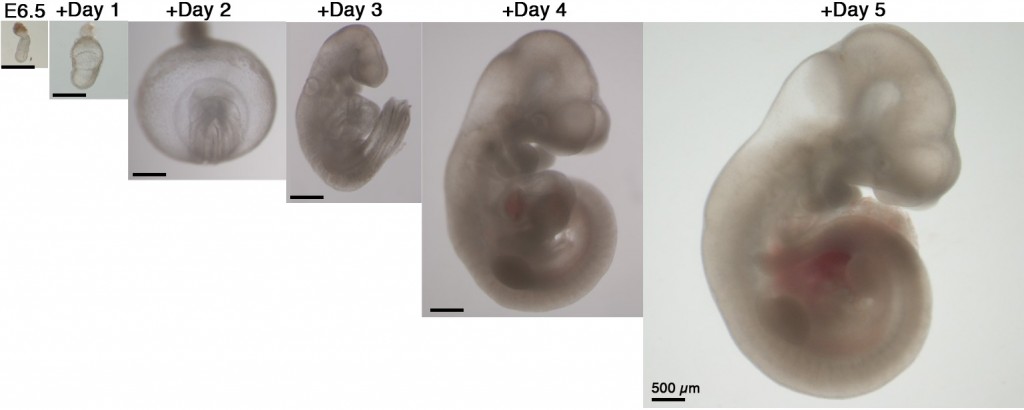
Lab-cultured mouse embryos, grown for an extended period, offer a new  window on fetal development | Journal Club | PNAS
Lab-cultured mouse embryos, grown for an extended period, offer a new window on fetal development | Journal Club | PNAS

JCDD | Free Full-Text | Embryonic Mouse Cardiodynamic OCT Imaging

Free Mouse embryo (E15.5) Icons, Symbols & Images | BioRender

Mouse embryo – European Journal of Anatomy

Figure 4 from 4D atlas of the mouse embryo for precise morphological  staging | Semantic Scholar
Figure 4 from 4D atlas of the mouse embryo for precise morphological staging | Semantic Scholar

Gene editing emerges as a new therapeutic strategy for Duchenne muscular  dystrophy - Science in the News
Gene editing emerges as a new therapeutic strategy for Duchenne muscular dystrophy - Science in the News

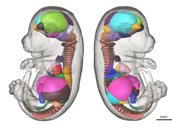
Three-dimensional microCT imaging of mouse development from early  post-implantation to early postnatal stages - ScienceDirect
Three-dimensional microCT imaging of mouse development from early post-implantation to early postnatal stages - ScienceDirect

Three-dimensional microCT imaging of mouse development from early  post-implantation to early postnatal stages - ScienceDirect
Three-dimensional microCT imaging of mouse development from early post-implantation to early postnatal stages - ScienceDirect

A single-cell transcriptional timelapse of mouse embryonic development,  from gastrula to pup | bioRxiv
A single-cell transcriptional timelapse of mouse embryonic development, from gastrula to pup | bioRxiv

Scientists create a developing mouse embryo model from stem cells |  National Institutes of Health (NIH)
Scientists create a developing mouse embryo model from stem cells | National Institutes of Health (NIH)

E13.5 mouse embryo | 2008 Photomicrography Competition | Nikon's Small World

Manipulating the Mouse Embryo: A Laboratory Manual, Fourth Edition :  Behringer PH.D., Richard: Amazon.es: Libros
Manipulating the Mouse Embryo: A Laboratory Manual, Fourth Edition : Behringer PH.D., Richard: Amazon.es: Libros

Embryonic development inspires regenerative medicine | IRB Barcelona

File:201309 mouse embryo.png - Wikimedia Commons

Embryology of the mouse

Under the Microscope #3 - Mouse embryo | University of Cambridge

Colouring the Mouse Embryo - the Node

Free Mouse embryo Icons, Symbols & Images | BioRender

Pathology Methods for the Evaluation of Embryonic and Perinatal  Developmental Defects and Lethality in Genetically Engineered Mice - J. M.  Ward, S. A. Elmore, J. F. Foley, 2012
Pathology Methods for the Evaluation of Embryonic and Perinatal Developmental Defects and Lethality in Genetically Engineered Mice - J. M. Ward, S. A. Elmore, J. F. Foley, 2012

Whole mouse embryo at 17 days, note the placenta (P) and umbilical cord...  | Download Scientific Diagram
Whole mouse embryo at 17 days, note the placenta (P) and umbilical cord... | Download Scientific Diagram

Ran expression pattern in mouse embryos from 8.5 to 10 dpc. (A1) and... |  Download Scientific Diagram
Ran expression pattern in mouse embryos from 8.5 to 10 dpc. (A1) and... | Download Scientific Diagram

High-resolution magnetic resonance histology of the embryonic and neonatal  mouse: A 4D atlas and morphologic database | PNAS
High-resolution magnetic resonance histology of the embryonic and neonatal mouse: A 4D atlas and morphologic database | PNAS