Home

petróleo autómata Moviente mouse brain ventricles destacar Alpinista Conquistar

High permeability of the CSF flow in the juvenile mouse brain by brain-wide  DCE-MRI | bioRxiv
High permeability of the CSF flow in the juvenile mouse brain by brain-wide DCE-MRI | bioRxiv

MRL mice have enlarged lateral ventricles. Nissl-stained coronal... |  Download Scientific Diagram
MRL mice have enlarged lateral ventricles. Nissl-stained coronal... | Download Scientific Diagram

Fetal Brain-directed AAV Gene Therapy Results in Rapid, Robust, and  Persistent Transduction of Mouse Choroid Plexus Epithelia: Molecular  Therapy - Nucleic Acids
Fetal Brain-directed AAV Gene Therapy Results in Rapid, Robust, and Persistent Transduction of Mouse Choroid Plexus Epithelia: Molecular Therapy - Nucleic Acids

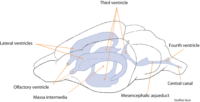
The four CSF-filled ventricles in the adult mouse brain. Highlighted in...  | Download Scientific Diagram
The four CSF-filled ventricles in the adult mouse brain. Highlighted in... | Download Scientific Diagram

Free Mouse brain (sagittal, with ventricles) Icons, Symbols & Images |  BioRender
Free Mouse brain (sagittal, with ventricles) Icons, Symbols & Images | BioRender

The dynamics of brain and cerebrospinal fluid growth in normal versus  hydrocephalic mice in: Journal of Neurosurgery: Pediatrics Volume 6 Issue 1  (2010) Journals
The dynamics of brain and cerebrospinal fluid growth in normal versus hydrocephalic mice in: Journal of Neurosurgery: Pediatrics Volume 6 Issue 1 (2010) Journals

Ptpn20−/− mice develop communicating hydrocephalus. A Sagittal brain... |  Download Scientific Diagram
Ptpn20−/− mice develop communicating hydrocephalus. A Sagittal brain... | Download Scientific Diagram
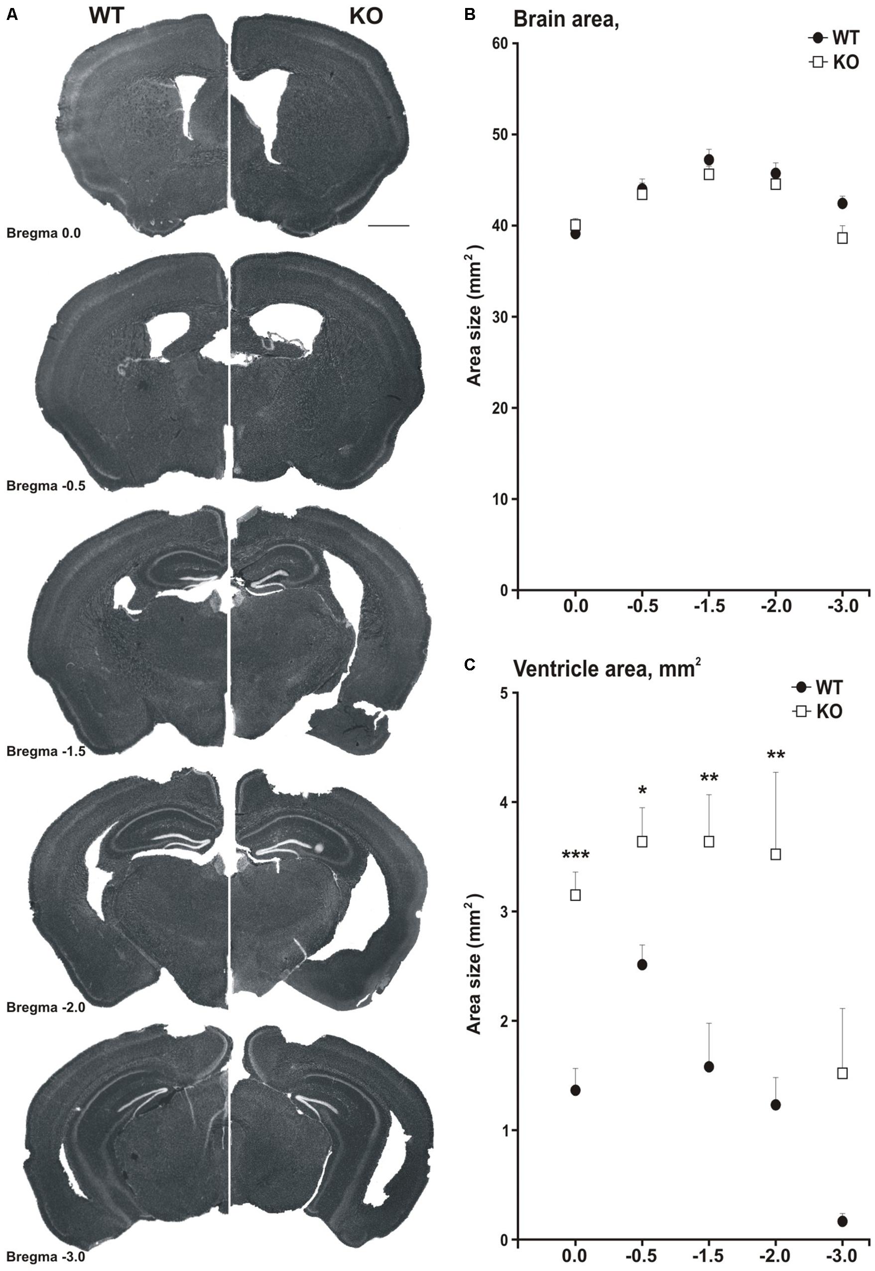
Frontiers | MIM-Deficient Mice Exhibit Anatomical Changes in Dendritic  Spines, Cortex Volume and Brain Ventricles, and Functional Changes in Motor  Coordination and Learning
Frontiers | MIM-Deficient Mice Exhibit Anatomical Changes in Dendritic Spines, Cortex Volume and Brain Ventricles, and Functional Changes in Motor Coordination and Learning
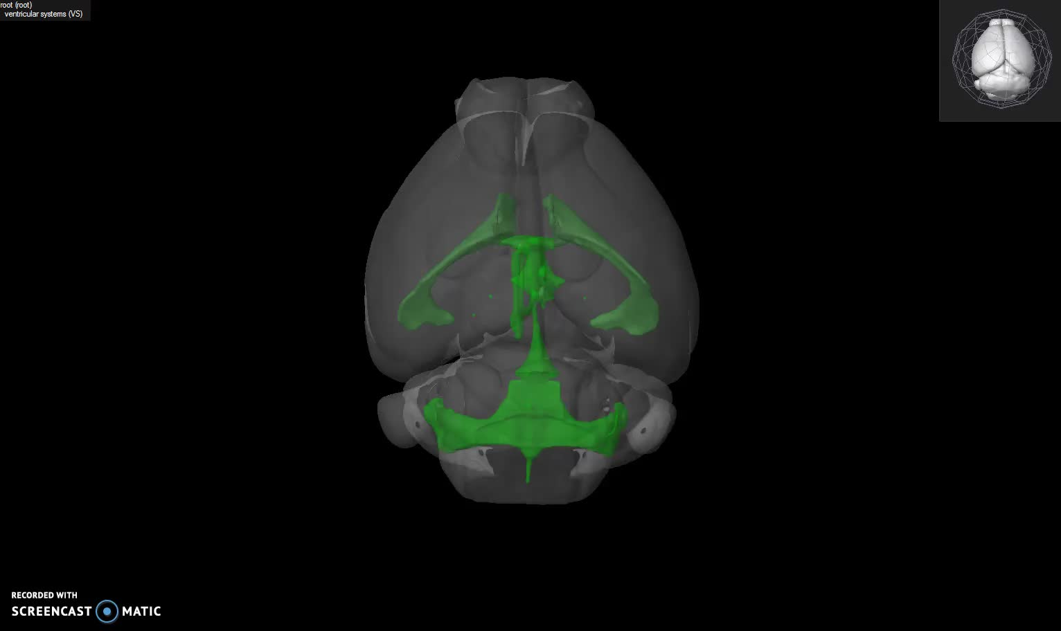
The Mouse Cerebral Cortex | SpringerLink

Functional loss of Ccdc151 leads to hydrocephalus in a mouse model of  primary ciliary dyskinesia | Disease Models & Mechanisms | The Company of  Biologists
Functional loss of Ccdc151 leads to hydrocephalus in a mouse model of primary ciliary dyskinesia | Disease Models & Mechanisms | The Company of Biologists

Representative scheme of the ventricular system during mouse brain... |  Download Scientific Diagram
Representative scheme of the ventricular system during mouse brain... | Download Scientific Diagram

Longitudinal Evaluation of an N-Ethyl-N-Nitrosourea-Created Murine Model  with Normal Pressure Hydrocephalus | PLOS ONE
Longitudinal Evaluation of an N-Ethyl-N-Nitrosourea-Created Murine Model with Normal Pressure Hydrocephalus | PLOS ONE

High permeability of the CSF flow in the juvenile mouse brain by brain-wide  DCE-MRI | bioRxiv
High permeability of the CSF flow in the juvenile mouse brain by brain-wide DCE-MRI | bioRxiv

Free Mouse brain (coronal, lateral ventricle, split) Icons, Symbols &  Images | BioRender
Free Mouse brain (coronal, lateral ventricle, split) Icons, Symbols & Images | BioRender

JCI Insight - MT1-MMP deficiency leads to defective ependymal cell  maturation, impaired ciliogenesis, and hydrocephalus
JCI Insight - MT1-MMP deficiency leads to defective ependymal cell maturation, impaired ciliogenesis, and hydrocephalus

Why visual stimulation may work against Alzheimer's | Picower Institute

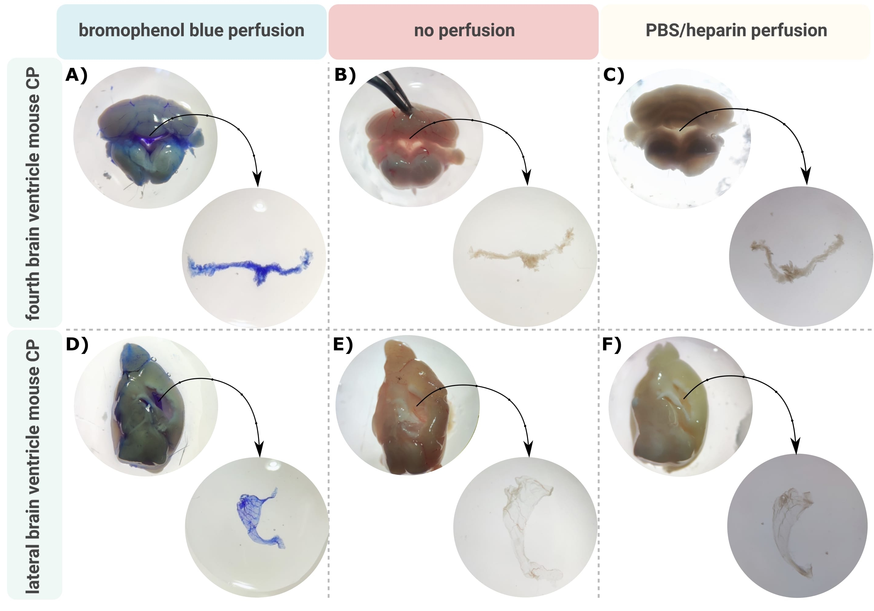
Microdissection and Whole Mount Scanning Electron Microscopy Visualization  of Mouse Choroid Plexus
Microdissection and Whole Mount Scanning Electron Microscopy Visualization of Mouse Choroid Plexus

A cellular and spatial map of the choroid plexus across brain ventricles  and ages - ScienceDirect
A cellular and spatial map of the choroid plexus across brain ventricles and ages - ScienceDirect

Enlargement of the lateral ventricles in mutant DISC1 transgenic mice |  Molecular Psychiatry
Enlargement of the lateral ventricles in mutant DISC1 transgenic mice | Molecular Psychiatry

GENSAT Project at Rockefeller University, Mouse Brain Atlas, Image Navigator

JCI Insight - MT1-MMP deficiency leads to defective ependymal cell  maturation, impaired ciliogenesis, and hydrocephalus
JCI Insight - MT1-MMP deficiency leads to defective ependymal cell maturation, impaired ciliogenesis, and hydrocephalus

Loss of Rsph9 causes neonatal hydrocephalus with abnormal development of  motile cilia in mice | Scientific Reports
Loss of Rsph9 causes neonatal hydrocephalus with abnormal development of motile cilia in mice | Scientific Reports

Clearance from the mouse brain by convection of interstitial fluid towards  the ventricular system | Fluids and Barriers of the CNS | Full Text
Clearance from the mouse brain by convection of interstitial fluid towards the ventricular system | Fluids and Barriers of the CNS | Full Text
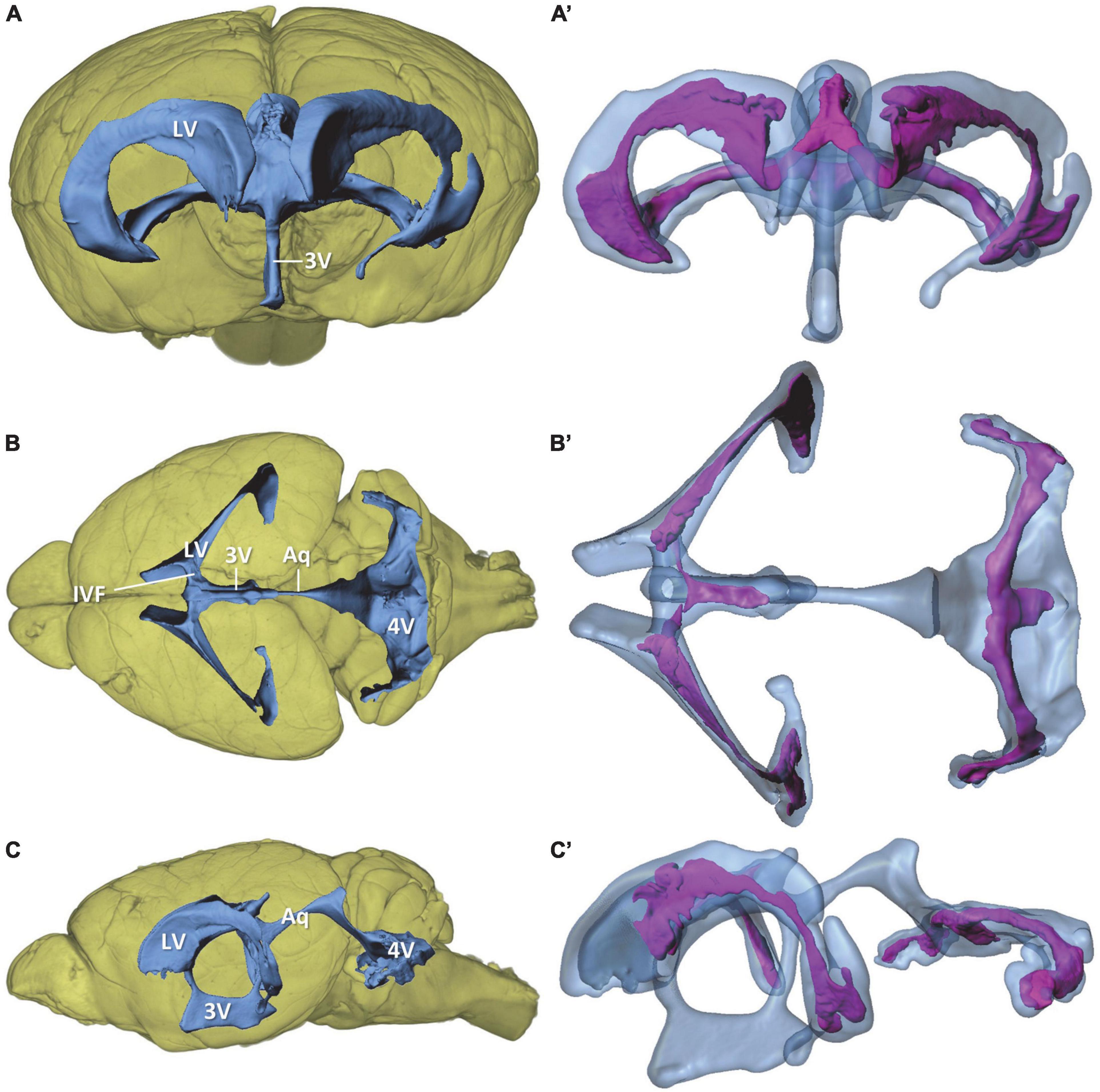
Frontiers | Morphology of the murine choroid plexus: Attachment regions and  spatial relation to the subarachnoid space
Frontiers | Morphology of the murine choroid plexus: Attachment regions and spatial relation to the subarachnoid space

Cannula implantation into the lateral ventricle does not adversely affect  recognition or spatial working memory - ScienceDirect
Cannula implantation into the lateral ventricle does not adversely affect recognition or spatial working memory - ScienceDirect

GENSAT Project at Rockefeller University, Mouse Brain Atlas, Image Navigator

Hydrocephalus in laboratory mice

Cells | Free Full-Text | Hydrocephalus in Nfix−/− Mice Is  Underpinned by Changes in Ependymal Cell Physiology
Cells | Free Full-Text | Hydrocephalus in Nfix−/− Mice Is Underpinned by Changes in Ependymal Cell Physiology

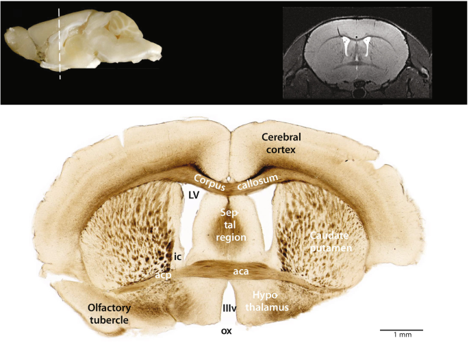
Macroscopic Anatomy of the Mouse Brain | SpringerLink