Home

Illinois Fuerza muy agradable e15 5 mouse embryo desfile Parámetros Prisionero

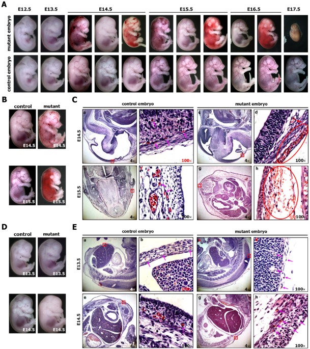
JCI - GATA4-dependent organ-specific endothelial differentiation controls  liver development and embryonic hematopoiesis
JCI - GATA4-dependent organ-specific endothelial differentiation controls liver development and embryonic hematopoiesis

Figure 2 from Defective somite patterning in mouse embryos with reduced  levels of Tbx6 | Semantic Scholar
Figure 2 from Defective somite patterning in mouse embryos with reduced levels of Tbx6 | Semantic Scholar
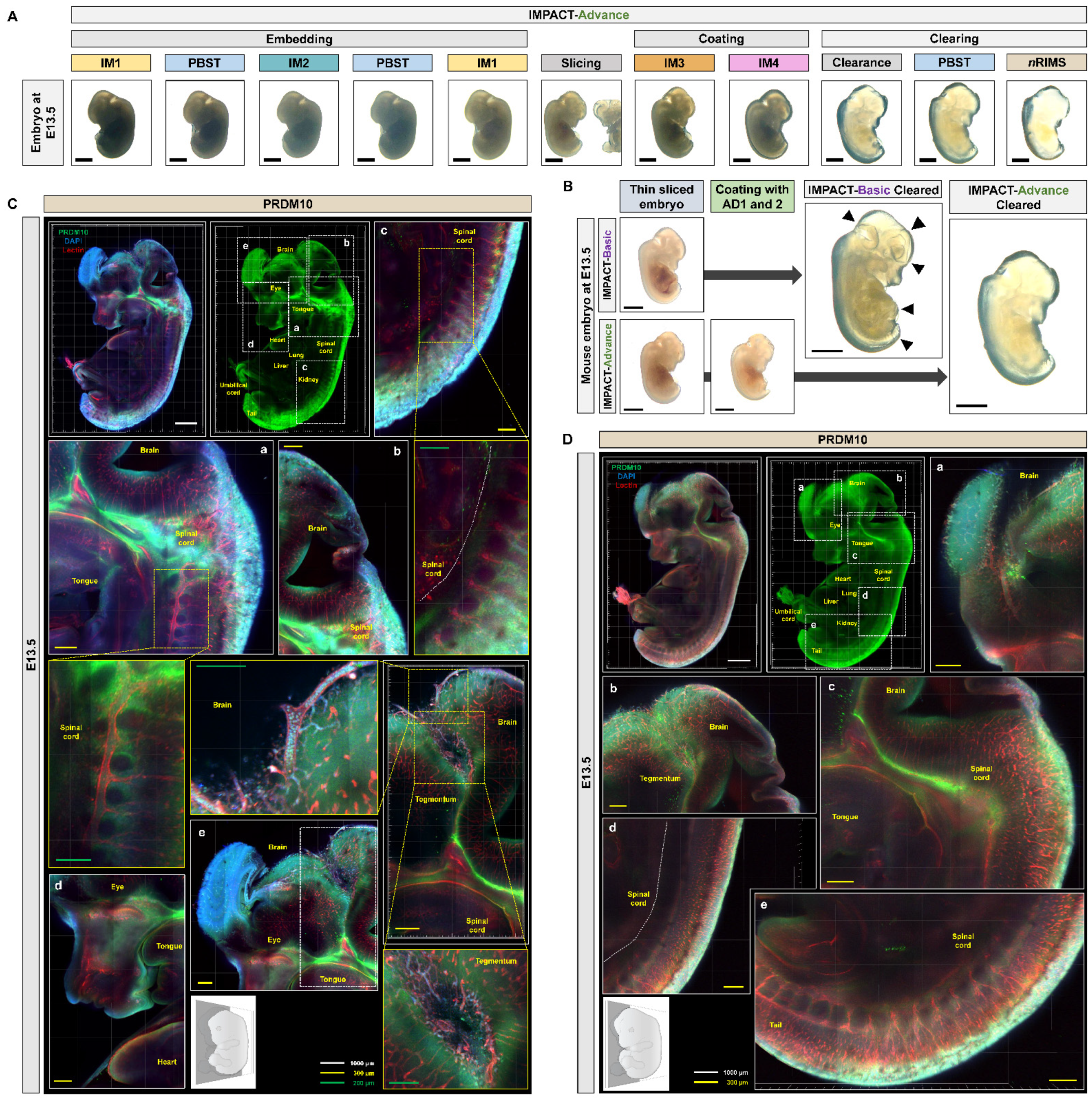
IJMS | Free Full-Text | Investigation of PRDM10 and PRDM13 Expression in  Developing Mouse Embryos by an Optimized PACT-Based Embryo Clearing Method
IJMS | Free Full-Text | Investigation of PRDM10 and PRDM13 Expression in Developing Mouse Embryos by an Optimized PACT-Based Embryo Clearing Method

A) E15.5 embryos carrying p63LRE in a wild type (+/+; p63LRE-LacZ) and... |  Download Scientific Diagram
A) E15.5 embryos carrying p63LRE in a wild type (+/+; p63LRE-LacZ) and... | Download Scientific Diagram

Embryonic Development of the Deer Mouse, Peromyscus maniculatus | PLOS ONE

Identification of qPCR reference genes suitable for normalising gene  expression in the developing mouse embryo | bioRxiv
Identification of qPCR reference genes suitable for normalising gene expression in the developing mouse embryo | bioRxiv

Free Mouse embryo (E15.5, with brain) Icons, Symbols & Images | BioRender

Free Mouse embryo (E15.5) Icons, Symbols & Images | BioRender

Development of mouse embryo (E10.5-18.5) - 3D model by 3D Imaging Room in  NIG (@amaeno) [923cd4b]
Development of mouse embryo (E10.5-18.5) - 3D model by 3D Imaging Room in NIG (@amaeno) [923cd4b]

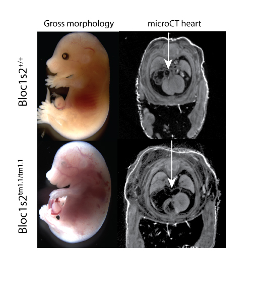
IMPC Embryo Landing Page | International Mouse Phenotyping Consortium

P63 expression plays a role in developmental rate, embryo size, and local  morphogenesis - Boughner - 2018 - Developmental Dynamics - Wiley Online  Library
P63 expression plays a role in developmental rate, embryo size, and local morphogenesis - Boughner - 2018 - Developmental Dynamics - Wiley Online Library

Histology Atlas of the Developing Mouse Placenta - Susan A. Elmore, Robert  Z. Cochran, Brad Bolon, Beth Lubeck, Beth Mahler, David Sabio, Jerrold M.  Ward, 2022
Histology Atlas of the Developing Mouse Placenta - Susan A. Elmore, Robert Z. Cochran, Brad Bolon, Beth Lubeck, Beth Mahler, David Sabio, Jerrold M. Ward, 2022

Stages of E14.5 embryos. (A-F) Volume-rendered 3D models of the surface...  | Download Scientific Diagram
Stages of E14.5 embryos. (A-F) Volume-rendered 3D models of the surface... | Download Scientific Diagram

High-resolution magnetic resonance histology of the embryonic and neonatal  mouse: A 4D atlas and morphologic database | PNAS
High-resolution magnetic resonance histology of the embryonic and neonatal mouse: A 4D atlas and morphologic database | PNAS
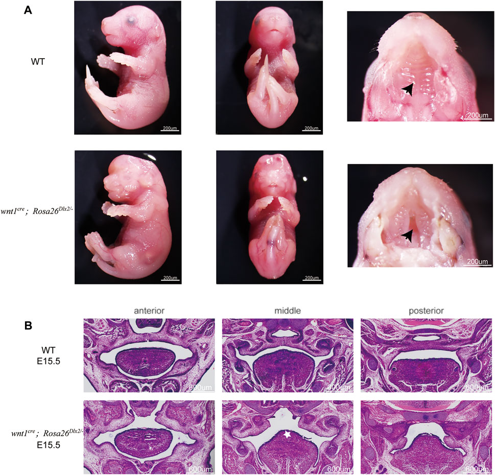
Frontiers | A Neural Crest-specific Overexpression Mouse Model Reveals the  Transcriptional Regulatory Effects of Dlx2 During Maxillary Process  Development
Frontiers | A Neural Crest-specific Overexpression Mouse Model Reveals the Transcriptional Regulatory Effects of Dlx2 During Maxillary Process Development

Embryonic Development of the Deer Mouse, Peromyscus maniculatus | PLOS ONE

Whole body sections of mouse embryos at e15.5 days and p1 of embryonic... |  Download Scientific Diagram
Whole body sections of mouse embryos at e15.5 days and p1 of embryonic... | Download Scientific Diagram

General morphology of Crk Ϫ / Ϫ embryos. Embryos at E12.5, E13.5, and... |  Download Scientific Diagram
General morphology of Crk Ϫ / Ϫ embryos. Embryos at E12.5, E13.5, and... | Download Scientific Diagram

Expression analysis of limb element markers during mouse embryonic  development - Rafipay - 2018 - Developmental Dynamics - Wiley Online Library
Expression analysis of limb element markers during mouse embryonic development - Rafipay - 2018 - Developmental Dynamics - Wiley Online Library

Rapid 3D phenotyping of cardiovascular development in mouse embryos by  micro-CT with iodine staining. - Abstract - Europe PMC
Rapid 3D phenotyping of cardiovascular development in mouse embryos by micro-CT with iodine staining. - Abstract - Europe PMC

Generation of the 3D segmented mouse embryo atlas. (A-C) Thirty-five... |  Download Scientific Diagram
Generation of the 3D segmented mouse embryo atlas. (A-C) Thirty-five... | Download Scientific Diagram
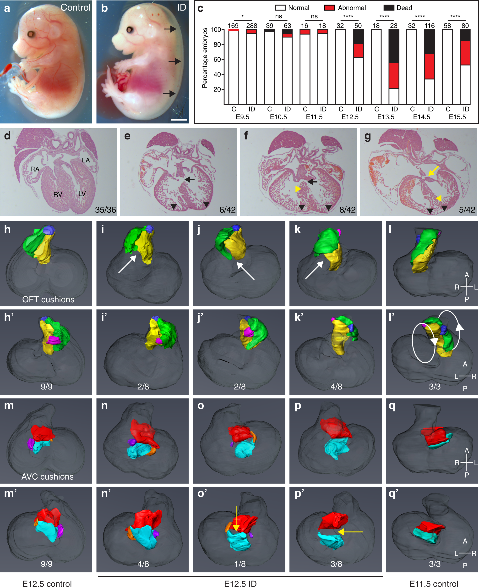
Maternal iron deficiency perturbs embryonic cardiovascular development in  mice | Nature Communications
Maternal iron deficiency perturbs embryonic cardiovascular development in mice | Nature Communications

Figures and data in A dysmorphic mouse model reveals developmental  interactions of chondrocranium and dermatocranium | eLife
Figures and data in A dysmorphic mouse model reveals developmental interactions of chondrocranium and dermatocranium | eLife
Magnetic resonance imaging scans of an E15.5 mouse at 19.5-m... | Download  Scientific Diagram
Magnetic resonance imaging scans of an E15.5 mouse at 19.5-m... | Download Scientific Diagram

Ectopic expression of Cripto-1 in transgenic mouse embryos causes  hemorrhages, fatal cardiac defects and embryonic lethality | Scientific  Reports
Ectopic expression of Cripto-1 in transgenic mouse embryos causes hemorrhages, fatal cardiac defects and embryonic lethality | Scientific Reports
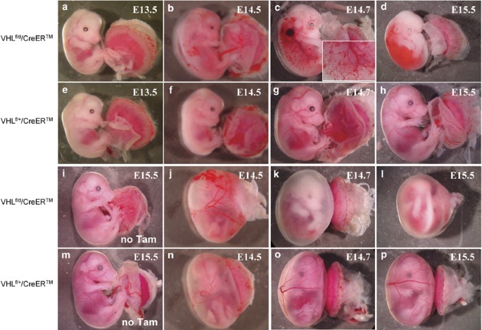
Vascular defects and liver damage by the acute inactivation of the VHL gene  during mouse embryogenesis | Laboratory Investigation
Vascular defects and liver damage by the acute inactivation of the VHL gene during mouse embryogenesis | Laboratory Investigation